Welcome
Our group uses numerical models to better understand and predict high-impact weather, including hurricanes and severe storms.
Our Research…
Graduate Student Opportunities
There are currently no prospective graduate student opportunities. Please click below to learn more about past opportunities.
More Details…
News & Awards
January 13, 2024 – Congratulations to Ari Tickner for successfully defending his M.S. thesis yesterday morning!
More Updates…
Our Group
We take pride in fostering supportive relationships in and beyond the academic environment. Check out the About Us page to learn more about us and our research! Group alumni have gone on to employment in research, operations, and the private sector.

Kade Barkas
(they/them)

Dillon Blount

Kathryn Boyle

Collin DeYoung

Brian Foster
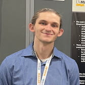
Drew Hickok